Single Extracellular Vesicle Protein Analysis by Immuno-ddPCR
How antibody-oligonucleotide conjugates turn digital PCR into a single-EV protein assay
Key takeaways
- Immuno-droplet digital PCR (iddPCR) repurposes ddPCR for protein analysis by using antibody-oligonucleotide conjugates to convert a single-EV protein-binding event into an amplifiable DNA barcode counted across thousands of droplet partitions.
- The reported assay reached a 38 EV/µL limit of detection, a three-order-of-magnitude dynamic range, and a strong correlation with nanoparticle tracking analysis (R² = 0.99).
- Multiplexed readout using FAM, HEX, and Cy5 enabled single-vesicle phenotyping of markers including PD-L1, GZMB, and TCF7, exposing PD-L1 heterogeneity across tumor-derived EVs (MC38 20.3%, B16 18.6%, KP1.9 8.6%).
- Each 65 bp barcode carried a unique 20 bp recognition sequence flanked by a universal PCR scaffold, so conjugate quality and oligo design are core determinants of assay performance rather than secondary details.
Digital droplet PCR (ddPCR) was developed for high-precision, absolute quantification of nucleic acid targets by partitioning a sample into discrete water-in-oil droplets, amplifying targets within each partition, and counting positive versus negative droplets without relying on a standard curve. In immuno-droplet digital PCR (iddPCR), that same digital readout is repurposed for protein analysis by using antibody-oligonucleotide conjugates to convert a protein-binding event into a DNA barcode that can be amplified and counted.
For extracellular vesicles (EVs), that molecular logic is especially useful. EVs are small, carry low amounts of protein cargo, and are compositionally heterogeneous. Bulk EV analysis can therefore miss biologically important subpopulations, whereas single-EV methods are designed to resolve heterogeneity directly at particle level. iddPCR was developed to address exactly that problem by enabling multiplexed single-EV protein profiling.
Why This Matters
Single-EV analysis is scientifically important because vesicle populations can be more heterogeneous than the parental cells from which they are shed. When all vesicles are analyzed together, low-abundance or phenotypically distinct EV subpopulations can be masked by the bulk signal. That is a major limitation for biomarker discovery, liquid biopsy, and translational workflows that depend on resolving rare vesicle populations rather than measuring only averaged abundance.
From an AbOliGo perspective, this is also a strong example of how antibody oligonucleotide conjugation extends beyond imaging and sequencing. In iddPCR, the oligo is not just a passive label. It becomes the molecular bridge between protein recognition and nucleic acid amplification, placing this method in the same wider AOC family as immuno-PCR and other DNA-enabled protein assays.
How the iddPCR Workflow Operates
The workflow begins by labeling EVs with antibody-DNA conjugates. These reagents bind proteins of interest on the EV surface and introduce DNA barcodes that can later be amplified by PCR. Free, unbound antibody-DNA conjugates are then removed by size exclusion chromatography (SEC) before droplet generation.
Single EVs are then stochastically incorporated into droplets together with PCR master mix using droplet microfluidics. After thermal cycling, droplets containing EV-associated barcodes become fluorescent and can be scored by imaging. This enables protein detection at the level of single droplets rather than bulk fluorescence across the full sample.
This droplet logic is what gives the method its analytical power. Standard ddPCR partitions a reaction into about 20,000 droplets and determines concentration by counting positive and negative partitions. iddPCR uses that same partitioning principle, but with the DNA signal now originating from an oligo-conjugated antibody bound to an EV-associated protein target.
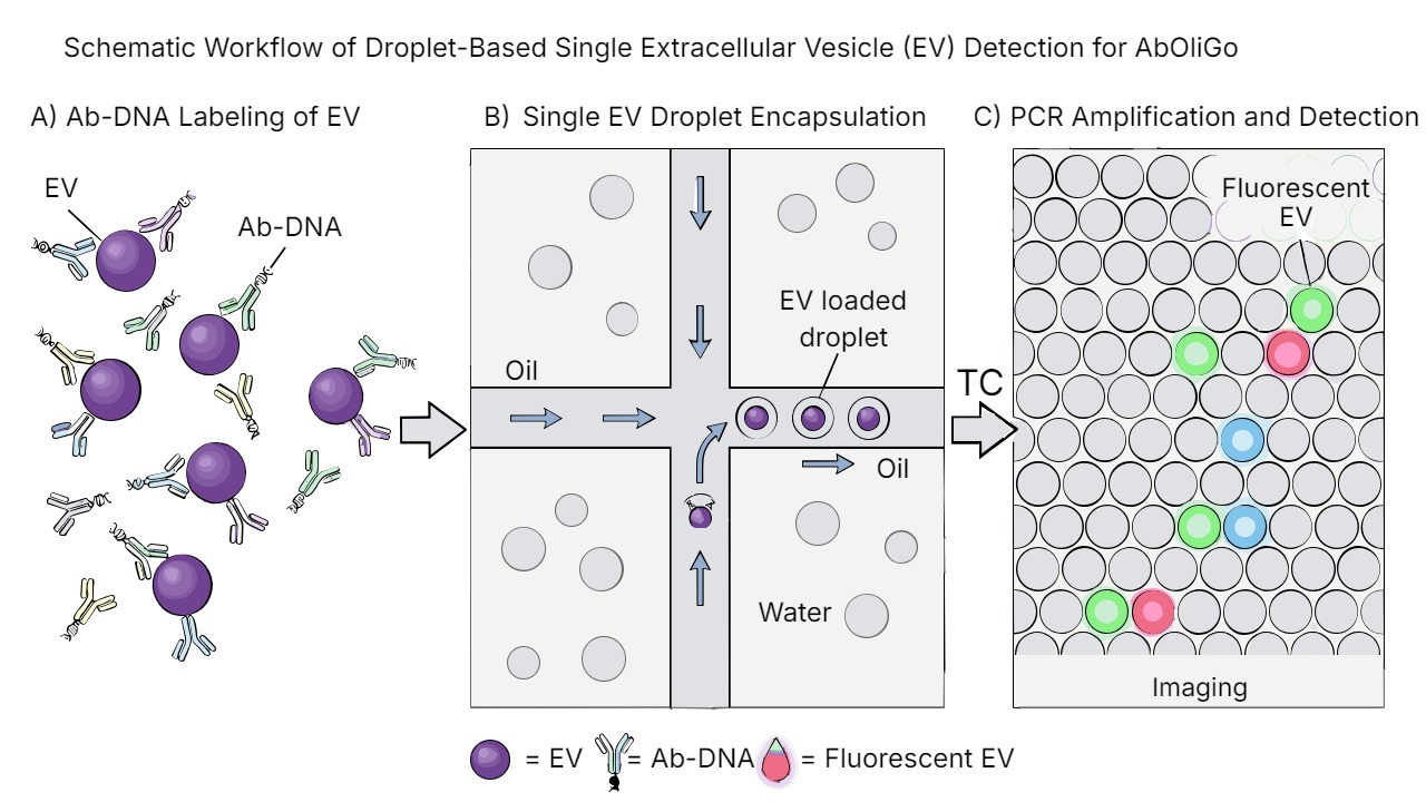
Single-EV detection in three steps. A. EVs are labeled with antibody-DNA (Ab-DNA) conjugates, and unbound conjugates are removed by size exclusion chromatography. B. Single EVs are encapsulated in droplets containing the EV and PCR master mix. C. After thermal cycling (TC), EVs labeled with the targets of interest amplify and fluoresce, and are analyzed by imaging.
Barcode and Conjugate Design
Each barcode was 65 bp in length and contained a unique 20 bp recognition sequence flanked by a universal scaffold for PCR amplification. Forward and reverse primers were designed to amplify the barcode, and fluorochrome-quencher probe pairs were used for multiplex readout. This is a good illustration of why oligonucleotide conjugation quality matters in advanced assays: the antibody provides target specificity, but the oligo defines how the signal is encoded, amplified, and decoded.
That same theme runs through AbOliGo's resources on approaches for making antibody-oligonucleotide conjugates and fine tuning AOCs, where conjugation strategy, degree of labeling, and assay compatibility are treated as core determinants of performance rather than secondary details.
Multiplexing Readout
The method was designed for multiplex protein detection at single-vesicle level. The article describes three-target profiling using FAM, HEX, and Cy5 fluorophores, and shows multiplexed analysis of markers including PD-L1, GZMB, and TCF7 in mouse plasma EVs. The same framework was also used for duplex analysis of other EV-associated proteins.
Scientifically, this matters because multiplexing allows individual EV subpopulations to be identified by marker pattern rather than by one marker alone. That pushes the assay beyond simple detection and toward single-EV phenotyping, which is exactly where antibody-oligonucleotide conjugates become most valuable as programmable assay components.
Analytical Performance
The reported analytical performance was strong for a single-EV protein assay. The paper reports a limit of detection of 38 EV µL⁻¹, a working dynamic range of three orders of magnitude, and a strong linear correlation between nanoparticle tracking analysis and ddPCR measurements with R² = 0.99.
The method was also applied to PD-L1 profiling in tumor-derived EVs. At matched EV concentrations, the MC38 sample had the highest PD-L1-positive EV population at 20.3%, followed by B16 at 18.6% and KP1.9 at 8.6%. The paper highlights this as the first direct determination of the quantity and fraction of PD-L1 positivity in single EVs shed from parental tumor cells.
These are the kinds of distributions that bulk analysis can miss. For liquid biopsy and biomarker development, the practical value of iddPCR is therefore not only high sensitivity, but the ability to expose protein heterogeneity at single-vesicle level.
AbOliGo Knowledge Hub
Within our Knowledge Hub, iddPCR sits naturally alongside scientific techniques using antibody-oligonucleotide conjugates and single-exosome profiling by proximity barcoding assay. The readout differs (ddPCR here, sequencing in PBA), but the assay logic is shared: antibody binding is converted into nucleic acid information that can be amplified, resolved, and quantified with much higher sensitivity than a conventional protein-only assay.
For AbOliGo, that is the broader signal. High-quality antibody-oligonucleotide conjugates, robust antibody oligonucleotide conjugation, and well-controlled barcode design are often the rate-limiting reagents in next-generation protein assays. iddPCR is a clear example of how those reagents can unlock digital protein counting at single-particle level.
Summary
Immuno-ddPCR extends ddPCR from nucleic acid quantification into single extracellular vesicle protein analysis by using antibody-DNA conjugates as barcode-bearing affinity reagents. In the article, EVs were labeled with 65 bp DNA barcodes containing 20 bp recognition regions, encapsulated into droplets, and read out by fluorescent PCR amplification. The method achieved multiplexed single-EV protein profiling, a three-order dynamic range, a 38 EV µL⁻¹ limit of detection, and direct measurement of PD-L1 heterogeneity across tumor-derived EV populations.
This article makes iddPCR one of the clearest examples of how antibody-oligonucleotide conjugates can transform a highly sensitive DNA technology into a highly sensitive protein assay, particularly in settings where rare targets, limited material, and biological heterogeneity matter.
Abbreviation Table
| Abbreviation | Meaning |
|---|---|
| AOC | Antibody-oligonucleotide conjugate |
| ddPCR | Digital droplet polymerase chain reaction |
| iddPCR | Immuno-droplet digital polymerase chain reaction |
| EV | Extracellular vesicle |
| PCR | Polymerase chain reaction |
| SEC | Size exclusion chromatography |
| TCO | trans-Cyclooctene |
| Tz | Tetrazine |
| PD-L1 | Programmed death-ligand 1 |
| EGFR | Epidermal growth factor receptor |
| EpCAM | Epithelial cell adhesion molecule |
| GZMB | Granzyme B |
| TCF7 | Transcription factor 7 |
| FAM | Fluorescein-based fluorescent reporter dye |
| HEX | Hexachloro-fluorescein fluorescent reporter dye |
| Cy5 | Cyanine 5 fluorescent dye |
Related reading
- Scientific Techniques Using Antibody-Oligonucleotide Conjugates
- Single-Exosome Profiling by Proximity Barcoding Assay
- Approaches for Making Antibody-Oligonucleotide Conjugates
- Fine Tuning Your Antibody-Oligonucleotide Conjugates